
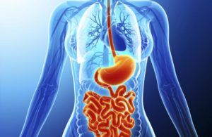
Gastroenterology refers to an area of medicine that focuses on the digestive system and its disorders. The Gastrointestinal/Digestive System (GI System) The GI system is the organ system responsible for processing food, extracting nutrients from it, and eliminating waste.
The GI system is divided into two sections:
The digestive tract – A muscular tube that extends from mouth to anus. It is also known as the alimentary canal. It includes the mouth, pharynx (throat), esophagus, stomach, small intestine, and large intestine.
The accessory organs – The teeth, tongue, salivary glands, liver, gallbladder, and pancreas.
There are many disorders and diseases that can affect the organs and tissues of the GI system, some of these include:
- Cancer (among the most common)
- Gastroesophageal reflux disease (GERD)
- Peptic ulcer disease (PUD)
- Gastritis
- Crohn’s disease
- Gluten sensitivity and celiac disease
- Inflammatory bowel disease (IBD)
- Irritable bowel syndrome (IBS)
- Constipation
- Hemorrhoids
Ultrasound is a very useful and effective tool for determining abnormalities and cancers of the GI system. It can be used to help diagnose pain or distention (enlargement) and evaluate all organs of the GI tract. It can easily detect abscesses, appendicitis, problems of the gallbladder (including inflammation and gallstones), pancreatitis, and any abnormalities in the size or shape of the GI organs.
Signs/symptoms that may indicate a problem within the GI system can include:
- Abdominal pain
- Changes in appetite
- Changes in urination, including too much urination, too little urination, or pain with urinating
- Constipation, diarrhea, or irregularity of bowel movements
- Jaundice (yellowing of the skin and eyes)
- Pain when lifting
- Rapid weight loss or weight gain
As with any medical problem, early detection and intervention is extremely important.
Ultrasound
Ultrasound is a safe, painless, non-invasive method of ruling out, or discovering the presence of disease using sound. In an ultrasound screening, a technician uses a small probe called a transducer and gel placed directly on the skin. High-frequency sound waves travel from the probe through the gel and into the body. The probe collects the sounds that bounce back, and a computer uses those sound waves to create an image. This safe, and effective tool can help with the diagnosis of multiple conditions related to the tissues and organs of the body.
Doctors can use ultrasound to diagnose conditions such as:
Tumors and cysts – An ultrasound can find dense areas of tissue such as tumors or cysts. It shows cysts differently from tumors to help your doctor make a diagnosis.
Cardiovascular issues – Ultrasound can be used to find narrowed blood vessels, blockages to blood flow, or weakened, ballooning areas of the arteries (aneurism).
Infections – Ultrasound can detect the increase in blood flow that often occurs in areas where infection is present.
Thyroid conditions – Ultrasound can be used to detect thyroid issues such as growths or abnormal activity.
Uterine fibroids – Detection of fibroids (non-cancerous growths in the uterus) and other conditions related to the female reproductive system show up on ultrasound. An ultrasound can help doctors find the source of pelvic pain and abnormal bleeding.
Monitor Pregnancy – Perhaps the most well-known use of ultrasound, a fetal ultrasound (sonogram) allows a healthcare provider to evaluate a baby’s growth and development and detect possible problems.
Abnormalities in size or shape of virtually any organ or tissue of the body Unlike CT scans and x-rays, there is no potentially harmful radiation exposure involved in an ultrasound. Because of its safe, effective, painless, non-invasive nature, ultrasound screenings should be a part of everyone’s regular physical examination schedule.
Please have your screening done as soon as possible.